Cerebral cavernoma, also known as cavernous angioma, is a malformation of the blood vessels that can cause problems in the brain and spinal cord.
It appears during brain development around different regions of the brain, and the abnormality that occurs, when several cavernomas form, is known as multiple cavernomatosis.
Most often, cavernomas do not produce symptoms, but there are also cases in which the person suffers seizures, vision problems, weakness and tiredness, headache, difficulty speaking and understanding others.
In more delicate situations, the patient suffers from nausea and vomiting, numbness on one side of the body, sudden and severe headaches, loss of vision or double vision, and difficulties in maintaining balance.
In any case, when the aforementioned signs and symptoms occur, the person must seek medical advice.
How to detect cavernomas?
There are different methods to diagnose them. Often times, they are evidenced in images of the brain made to diagnose other neurological diseases.
In the presence of symptoms that suggest the presence of a cavernoma, doctors usually request specific tests to confirm or rule out a malformation or to identify or rule out other related diseases.
We also resort to images of the brain that allow us to reveal hemorrhages or the emergence of new malformations. In this case, we can resort to:
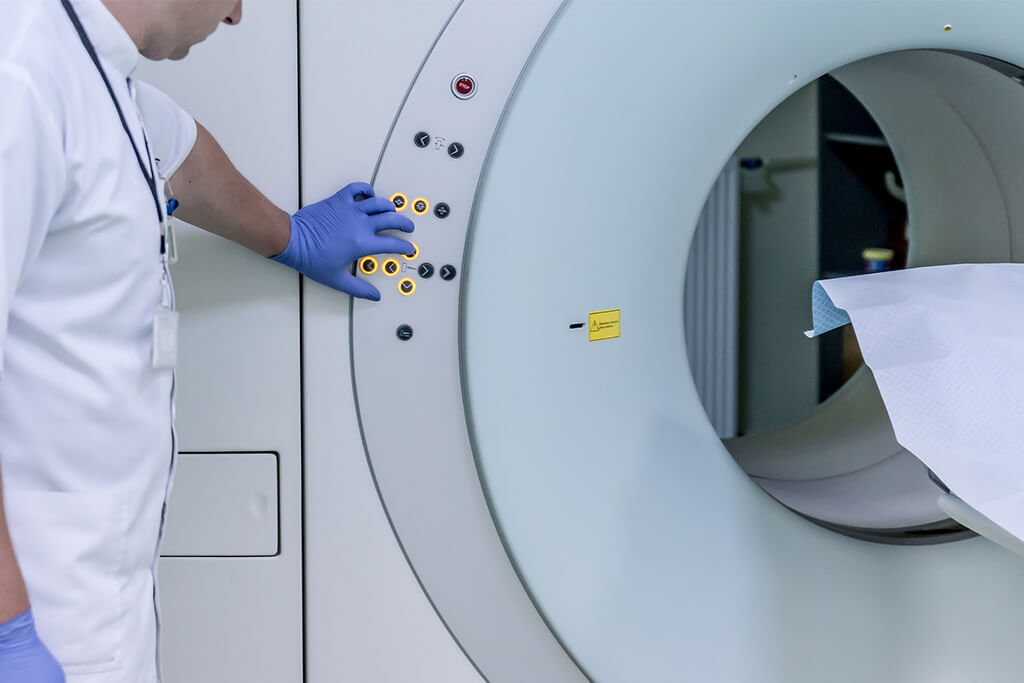
- Magnetic resonance imaging (MRI). It helps us to obtain a detailed image of the brain, the spinal column and the blood vessels of the brain. We also have magnetic resonance angiography (or MRI venography) in our favor, which consists of injecting a contrast dye into a vein in the arm to better see the blood vessels in the brain.
- Genetic analysis. We use them to identify the changes in genes or chromosomes associated with cavernous brain malformations, when the patient has a family history of the disease.
Vascular surgery
The most advanced technology allows us to give high precision treatment to cavernomas through minimally invasive surgeries that provide the patient with the additional benefit of recovering in less time than that required by open surgery.
In minimally invasive neurosurgery, we use an instrument called an endoscope, consisting of a small illuminated tubular camera that performs the function of a microscope.
With instruments that also do not require large incisions, we operate on certain areas of the brain, the base of the skull, or the spinal cord. It is a fast, precise and simple procedure, which allows a quick recovery, reduces pain and minimizes scars.
In endoscopic intracranial surgery, specifically, the instruments allow us to perform tumor biopsies, resection of colloid cysts, fenestrate cysts and treat hydrocephalus.
We are talking about operations that take just a few minutes and allow us to discharge the patient the next day.
Contact us, we are here to serve you.


